How Skin Color Works
Skin color or skin pigment is something that our students have typically loved learning about
and most of our students will tell us, yes, genetics plays a huge role in skin
color or skin pigment and even that the sun can influence how much pigment the skin produces
but what layer specifically is responsible for the skin pigment and more importantly,
what are some of those differences that you might see inside the skin
with somebody with lighter skin all the way up to darker skin and everywhere in between.
That answer might surprise you and you may have noticed that those with darker skin have a
greater difference in the pigment of the palm of their hand and the dorsal aspect of their hand,
we'll talk about why that is and even go into some sun damage and things of that nature
as well as think about this... if I were to remove the skin from a body,
yes, I do understand that sounds a little bit morbid but we are a cadaver lab,
but if I were to line up all the bodies we have in our lab in a row, remove the skin, and have
you look at those bodies, you couldn't tell me the difference between each body, meaning which body
had which pigment of skin prior to the removal of the skin. Kind of interesting to think about.
So, let's jump right into this.
♪ ♪
So, for us to fully appreciate skin color or skin pigment, we have to review the skin a
little bit. The skin belongs to a system called the integumentary system or the integument and
it's comprised of three main layers. So, let's take a look at the cadaver dissection that we
have here. This is a skin graft from the mid back and you can see this top layer or the superficial
layer of the integument is referred to as the epidermis. Epi just means upon, dermis means skin.
Now, what's really cool is if I flip up the edge here, you might think that the edge is the
epidermis but the majority of that edge that I'm running the pro bond is actually the next layer
down. It's that top paper thin layer right there that actually makes up the epidermis, so pretty
thin. And when I say paper thin, the majority of the epidermis throughout the whole body is about
0.1 to 0.15 millimeters in thickness. Pretty close to the thickness of a sheet of paper.
Now, there are some other areas of the epidermis that are much thicker like on the palm or on the
soles of the feet. That can range from 0.6 up to 4.5 millimeters in thickness. Now, you
may not think 4.5 millimeters is that thick. But remember, the majority of the body is 0.1 to 0.15
and if you compare 0.15 millimeters up to 4.5 millimeters, that's a 30 fold increase in
thickness on the palms and the soles, which we are definitely gonna discuss later on in this video.
But if we come back to this dissection here, the next layer down that I hinted to was the dermis
or the dermal tissue and that's the majority of the thickness of this dissection here.
The dermis is a dense connective tissue that provides strength from like tensile forces
like the skin being pulled apart, it can resist being pulled apart. Also has hair
follicles pushing down into it, glands, and even blood vessels but the next layer down,
if I flip this over, is referred to as the hypodermis or the subcutaneous layer.
Now, we've removed most of the hypodermis and the subcutaneous layer from these
other areas but you can see that yellowy tissue where we kept it in certain areas
and the hypodermis is made out of an adipose tissue. An adipose can range from relatively thin
all the way up to inches thick and that adipose layer or that subcutaneous layer also known
as the hypodermis cuz anatomists love naming things, it is for energy storage and insulation.
Now, I just wanna be clear here, the true skin is actually the epidermis and the dermis.
We add the hypodermis to complete the whole integumentary system. So,
it's kinda like the epidermis and dermis, the skin is a subset
of the whole integument but what are we here for? We wanna talk about where the pigment comes from.
What's interesting is if we talk about tattoos, the majority of tattoo ink or pigment gets
deposited into the dermis and we have a pretty cool tattoo video that will link that at the
end of the video if you wanna take a look at that later but the majority of ink like I mentioned or
pigment from tattooing is in the dermal tissue but there's a big difference from depositing
artificial ink or pigment versus creating your own pigment and how we create our pigment
or where we create it is specifically in that epidermis which is crazy to think about again,
because the majority that is only 1-. 15. millimeters in thickness.
So, all the color that we've - well, I shouldn't say all, but the majority of our skin pigment,
some of it is influenced by like blood supply and things like that
but the pigment itself, just in that top paper thin layer,
that we call the epidermis. So, obviously, we're gonna have to go a little bit deeper
into the epidermis to fully understand pigment formation or pigment production.
So, if we zoomed into that top edge of the epidermis here, again, it would look paper thin to
the naked eye but as we zoomed in, we would see that it is cell upon cells stacked on top each
other or multiple cell layers thick. The majority of the cells of the epidermis or at least 90%
of the cells that make up the epidermis are referred to as keratinocytes. Cyte
meaning cell, keratin because they produce protective or strong protein called keratin.
That keratin helps bind the cells together and in turn, helps protect us on the top layer from
friction and abrasion. But the keratinocytes don't produce the pigment. That's gonna be
the job of another cell. Now, remember, we're really zooming into this epidermis and this
epidermis can be further subdivided or what we'll see is there's sub layers.
The bottom row of cells of the epidermis is referred to as the stratum basale and again,
the majority of that's going to have keratinocytes but it will have intersperse
between the keratinocytes, another cell that's important called the melanocyte.
Cyte meaning cell, but because they produce melanin, they get called
melanocytes and melanin is that pigment that gives the color to the skin.
Now, because the melanocytes interact with the basale, the bottom sub layer here and the
next sub layer above, I have to mention some key points with those sub layers. The stratum basale,
those keratinocytes are mitotically active. Meaning, they constantly divide and a cell will
divide and get pushed up to another layer and when the next cell is copied, the cell that was here,
then moves up and a new one comes in its place and eventually, this continues to happen so
that one cell that was at one point in the stratum basale will eventually make it all
the way to the top of your epidermis and flake off and contribute to the dust of your house.
Now, the melanocytes are gonna be doing something to those cells in the basal layer
but they'll also interact with the cells in the sub layer above
called the stratum spinosum. Now, the stratum basale was only one cell layer thick
but the stratum spinosum is up to eight to 10 cell layers thick.
Now, some of you are geeky anatomous and wanna know why things get named the way they did.
Now, these cells in the spinosum when they were looked at under the microscope, they would
shrink down. But because they were attached to all the other surrounding keratinocytes,
even though the majority of the cell would shrink down, the places where it was still
attached to the other cells would be kind of pulled out attaching to each other
and look like little spines on the outside of the cell membrane hence,
the name. But those melanocytes, let's really dive into these things now.
These are really cool cells that we already know produce melanin. They have these cool
things called cytoplasmic extensions or they look like little arms that are coming out from them
that are kind of just extensions of their cell membrane. That they can contact
up to 30 different keratinocytes. Some of those in the stratum basale
and some of those in the stratum spinosum and what they're gonna do is deposit melanin into
those keratinocytes and that will give each keratinocytes a color or a pigment.
Now, how much melanin is deposited in there will dictate how dark the cell gets.
Now, what's crazy to think about is that 8% of the epidermis or 8% of the epidermal cells are
melanocytes regardless of your skin pigment. So, if you're lighter skin or darker skin, you have
about the same number of melanocytes. It's just with darker skin those melanocytes are more active
and produce more melanin kind of at that baseline rate and what I mean by a baseline rate is all
of us kind of have our baseline pigment or color without UV stimulation or stimulation from the sun
and that just gives us kind of, like I said, our baseline skin color.
But if we go out in the sun, let's pretend all of us go on this little beach gathering together
and we're out frolicking in the sun, you know, swimming in the water,
whatever we like to do on the beach and that sun is coming down. That will emulate the melanocytes
to produce even more melanin. So, all the cells that are in contact in contact with
those melanocytes are getting extra melanin in it because we - most of us have probably heard
melanin is protective. It's protecting our keratinocytes specifically the nucleus and
the DNA inside the nucleus from DNA damage and potential mutation and so, more UV light
exposure, more potential for DNA damage. So, melanocytes are let's produce more and protect.
Now, we know that in general, people with darker pigment are less at risk for things like skin
cancer and UV damage. They're just more protective compared to somebody who has lighter skin and
when we talk about getting a tan, whether you think it's a good thing or a bad thing, there's
plenty of evidence that shows fair-skinned people getting exposed to UV light are at a greater risk
for skin cancer but why don't you maintain the tan is a question people will get.
Well, we all went home for our beach trip and weren't stimulated by the sun as much.
Now, we only had that UV stimulation for that day and the cells in that stratum spinosum and
stratum basale only got extra melanin deposited. So, when the base alley creates more cells for us,
it pushes those cells that are darker pigment from the tan, if you will,
and eventually those darker keratinocytes will flake off.
And so for those of you who like to be tan all the time, you know that you have to constantly
get UV stimulation to maintain that tan. For those of you who are afraid of the sun like myself, you
try to tend to avoid that overstimulation of melanocytes by taking on you know, SPF 50.
So what about the differences in pigment on the palms of the hand
and the soles of the feet? Well, the answer to that lies in the additional sublayers
of the epidermis. Now, remember, we already talked about two sublayers of the epidermis,
the stratum basale and the stratum spinosum. We're gonna add a couple of more to help us
understand these differences in the palms and the soles but let me mention one thing
about the epidermis. The epidermis is an Avascular tissue. Meaning, it doesn't have
a blood supply. There's no room for blood vessels to penetrate between those cells.
Typically, we like cells to have a blood supply in order for them to stay alive but the dermis
underneath the epidermis where the epidermis plots down on top of, does have a blood supply. So,
cells in the bottom, like the cells of the stratum basale, those are the most mytodically active,
they're close enough to blood that they can continue to divide and get those nutrients
but as the cells get pushed up further and further away from the blood, they start to
flatten out and that's exactly what happens in our next sublayer that we're gonna talk about.
This is called the stratum granulosum and yes, those cells are flattening out but they're also
releasing these little granules full of lipids and these granules, you can kinda think of
these little sacs filled of lipids and oils, and they're releasing them outside of themselves. So,
essentially, coating themselves in oil. Now, why would these cells do that?
Well, if we think about the mortal enemy of oil, that is water. So, we're creating this
waterproofing barrier in the epidermis so water cannot pass freely in and out of the epidermis.
Kind of cool but admittedly, there's no difference between the palms and the soles
and everywhere else in the body, they just have this typical granulosum in those areas.
But when we get to the top top layer called the stratum corneum, those cells have fully
flattened out and are completely dead. You'll have a stratum corneum everywhere in your body.
And in some areas, it can be a couple cell layers thick but in the palms and in the soles,
it can be up to 50 cell layers thick contributing to that extra thickness here.
Now, think about light trying to penetrate through more cell layers
and then, you may have noticed one other thing,
I didn't mention another sublayer called the stratum lucidum. The stratum lucidum is in
between the granulosum and the corneum. The reason I didn't mention it initially is because you
only find that on the palms of hand in the soles of the feet and nowhere else throughout the body.
Now, lucidum refers to like being lucid or transparent or full of light
and again, producing an additional thickness to the palms and the soles. So,
you're adding all this extra thickness and makes it harder for light to penetrate
through and it kinda makes sense when you look at your palms or the soles of your feet.
Now, a couple other things to consider, most of us aren't exposing our palms to the sun
very often either. Just the natural position of how we hold our hands.
They don't get exposed quite as much and definitely the soles as you're
just walking down on the ground a lot of the time don't get nearly as much sun exposure.
That's the same essentially with everybody with lighter pigmented skin all the way up to darker.
The difference is those with darker pigments, it's just more noticeable because they have that
darker pigment everywhere else so that change is a little bit more dramatic or more noticeable.
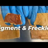
Now, like I mentioned at the beginning of the video, I have to touch a little bit on sun damage
and that's mostly because of our aesthetic students because whenever they come into the lab,
one of the first things they point out on this particular skin graph is they point to
all these little areas and they're like, "What? Sun damage?" And as you can see, they're pointing
out those areas of hyperpigmentation or other spots that a lot of us will refer to as freckles.
Now, there are two types of freckles. One is called Ephelides and the other is called
lentigines. Ephelides, that funny name there, is more of a inherited type freckle that you'll
see in really light-skinned people. It appears to just be a genetic thing.
Now, Lentigines is more of a sun damage or a sunburn freckle as some people will refer to it as
and that's because of over exposure to the sun and potential sun damage.
In these areas where you're seeing those freckles or those lentigines, that is a buildup of melanin
or little patches of melanin in those areas again, because of over exposure to the sun.
Now, I always say to people, yes, the evidence is pretty clear that light-skinned people are
more at risk for developing skin disorders and skin cancer because of UV exposure
but we'll let everybody decide how much sunscreen you wanna lube up with. You know,
I typically recommend throw the 50 on and go play in the water and frolic on the beach.
As always, thanks for watching everyone. If you wanna support the channel, you can subscribe,
smash the like button, even write some comments below and let us know what you thought of the
video as well as future videos that you might like to see. We've also got our T-shirts and our
anatomical artwork back there. We'll put the link in the description
and we'll see you in the next video.